
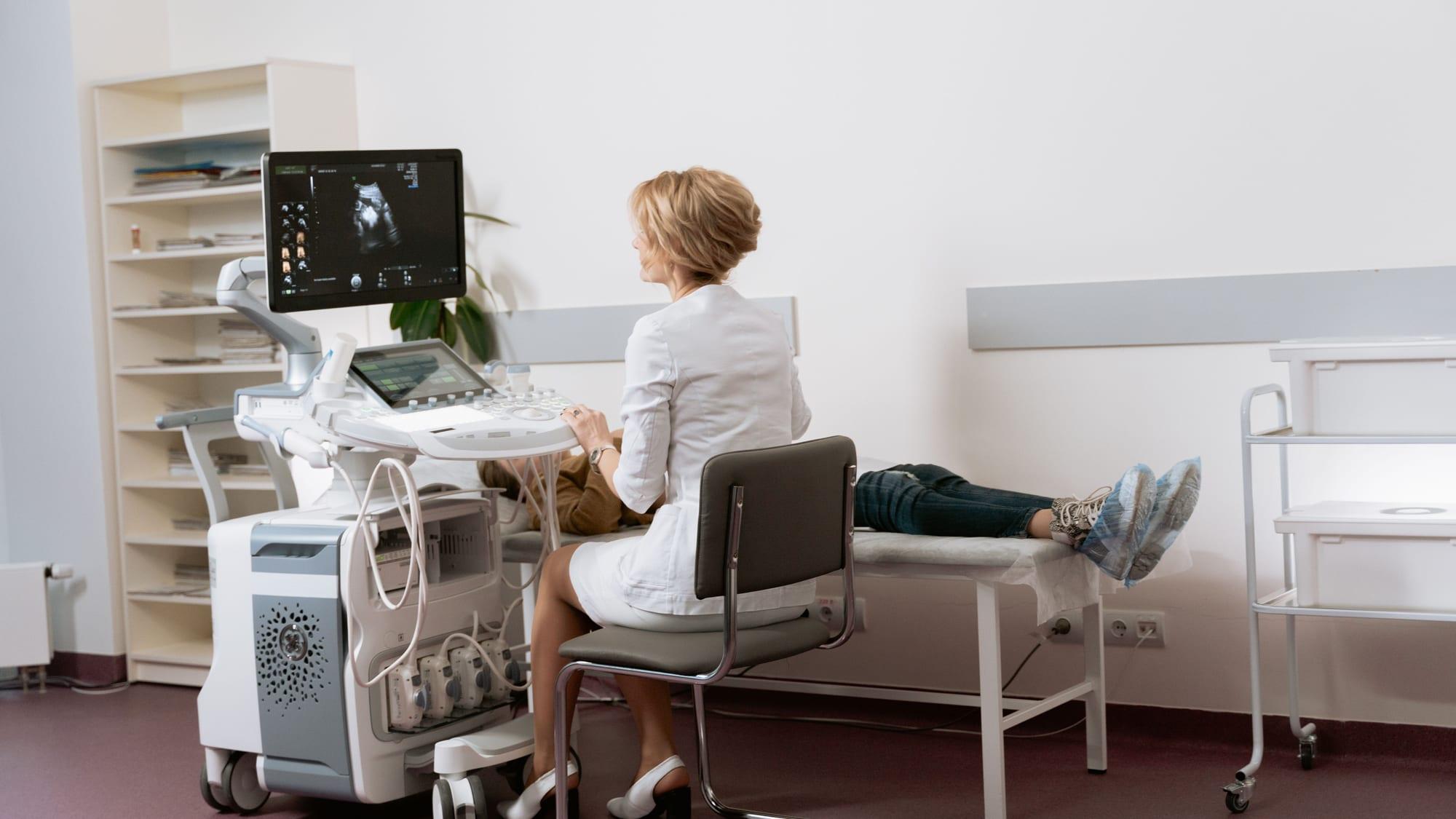
Echocardiography (echo) is a painless, non-invasive diagnostic method in which the heart is imaged using ultrasound waves. It provides detailed information about the size of the heart chambers, pumping strength, valve structure, and blood flow within the heart; it is frequently used in the investigation of complaints such as shortness of breath, a murmur, or palpitations, and in the follow-up of known heart failure or valve disease. This article covers when echo is requested, how it is performed, and how it differs from transesophageal echocardiography (TEE).
What Is Echocardiography (Echo)?
Echocardiography is a diagnostic method that provides real-time imaging of the heart using an ultrasound probe applied over the chest wall. No radiation is used during the procedure, and no needle or incision is required.
The standard form of the procedure is called transthoracic echocardiography and provides a comprehensive initial assessment of the heart's chambers, valves, and blood flow.
What Symptoms Prompt Echocardiography?
Because echocardiography directly addresses questions about the heart's structure and function, it is one of the methods frequently used in cardiology evaluation. The situations below are example scenarios in which this test may be considered; the final decision depends on the patient's individual examination.
- Increasing fatigue or shortness of breath during daily activities
- A heart murmur detected on examination
- Regular follow-up of known heart failure or valve disease
- A finding on ECG that may point to a structural problem
How Is Echo Performed?
The patient exposes the upper body and lies on the examination table on their left side; special preparation is generally not required. Ultrasound gel is applied to the chest area, and the probe is moved over the chest from different angles to image the heart chambers, valves, and blood flow from various windows.
What Does Echo Evaluate?
Echocardiography can evaluate the size and wall thickness of the heart chambers, the heart's pumping strength (ejection fraction), the structure and function of the heart valves (stenosis, regurgitation), blood flow within the heart, and whether there is fluid accumulation in the pericardium.
However, echo does not directly visualize narrowing in the coronary arteries; Coronary Angiography or CT Coronary Angiography is used for this purpose. It also does not continuously monitor the heart's electrical rhythm; ECG or Holter ECG is needed for rhythm assessment.
Difference Between Standard Echo and Transesophageal Echocardiography (TEE)
Standard (transthoracic) echocardiography is performed non-invasively over the chest wall and provides a first-line assessment of the heart's structure and function. In certain special situations where image quality is insufficient or more detailed examination is needed, Transesophageal Echocardiography (TEE), performed by advancing the probe into the esophagus, is considered as a separate procedure; TEE offers much more detailed imaging from a position closer to the heart.
Frequently asked questions
Echocardiography is a painless, non-invasive diagnostic method in which the heart is imaged using ultrasound waves. It provides detailed information about the heart chambers, pumping strength, heart valves, and blood flow.
It generally does not require special preparation. It is sufficient to wear clothing that can easily expose the upper body before the procedure; fasting is not necessary.
The procedure generally takes between 20 and 40 minutes; the duration can vary depending on the scope of the findings to be assessed.
No. During the procedure, no intervention is performed other than moving a probe over the chest; the ultrasound gel causes no discomfort other than a mild sensation of coolness.
No. Echocardiography evaluates the structure and function of the heart; it does not directly visualize narrowing in the coronary arteries. Methods such as Coronary Angiography or CT Coronary Angiography are used for this purpose.
No. Standard echocardiography is performed over the chest wall, while Transesophageal Echocardiography (TEE) is performed by advancing the probe into the esophagus, providing much more detailed imaging from a position closer to the heart.