
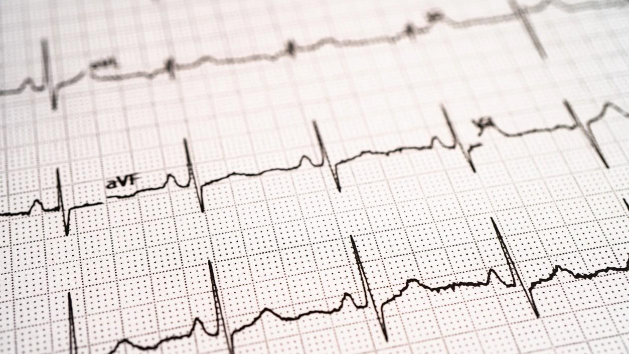
An ECG (electrocardiography) is a painless diagnostic method, completed within a few minutes, in which the electrical signals the heart generates with each beat are recorded through electrodes placed on the skin. It is used as one of the first-line tests in the investigation of complaints such as palpitations, chest pain, dizziness, or fainting, and provides rapid information about the heart's rhythm and electrical conduction pattern. This article covers when an ECG is requested, how it is performed, and how the result is evaluated.
What Is an ECG (Electrocardiography)?
An ECG is the recording, from the body surface, of the electrical signals the heart generates during each cycle of contraction and relaxation, using small electrodes placed on the chest, arms, and legs. The resulting recording is captured as a waveform showing the heart's beating rate, rhythm pattern, and the functioning of the electrical conduction system.
The procedure involves no needle or incision; the electrodes are attached to the skin with adhesive pads, and no electrical current is delivered to the patient — only the signals the heart itself generates are recorded. For this reason, the ECG is one of the most frequently used and safest cardiological screening methods in everyday practice.
What Complaints Prompt an ECG?
An ECG is a test used in the initial evaluation of many complaints that may be related to the heart. The situations below are example scenarios in which an ECG may be considered; the physician decides whether the test is needed based on the patient's history and examination findings.
- Palpitations, or feeling the heartbeat as irregular or fast
- Chest pain, or a feeling of pressure or tightness
- Unexplained dizziness, a feeling of fainting, or shortness of breath
- Routine health checkups and pre-operative evaluation
- Regular follow-up of a known heart condition
How Is an ECG Performed?
The patient lies down on the examination table, and the areas on the chest, arms, and legs where electrodes will be placed are exposed. After small adhesive electrodes are placed on the skin, the patient is asked to remain still and calm for a period ranging from a few seconds to a minute; the heart's electrical activity is recorded during this time.
No special preparation is required before the procedure; there is no need to fast or stop medications. Once the recording is complete, the electrodes are removed, and the result is generally evaluated by the physician and shared with the patient in the same session.
What Does an ECG Show, and What Doesn't It Show?
An ECG can screen for heart rate, rhythm pattern, whether there is delay or block in the electrical conduction system, and changes that may point to a prior heart attack or ischemia. In this respect, it is a fast and practical source of information in the initial evaluation of many heart-related complaints.
However, the ECG also has certain limitations. It does not visualize the heart's structure, meaning its chambers and valves; Echocardiography (echo) is needed for this. It may fail to capture intermittent and brief rhythm disturbances if they do not occur at the moment of recording; in such cases, longer-term recording methods such as Holter ECG are considered. It also does not show the heart's response during exercise; an Exercise (Stress) Test is used for this purpose.
How Is the Result Evaluated?
ECG findings are not evaluated in isolation but holistically, together with the patient's complaints, past cardiac history, and medications used. Whenever possible, the current recording is compared with the patient's previous ECG results, if available, to examine changes over time.
When a suspicious finding is detected on ECG, the physician evaluates whether further tests are needed, such as Echocardiography for structural assessment of the heart, Holter ECG if intermittent rhythm disturbance is suspected, or an Exercise (Stress) Test for findings that appear with exertion.
Frequently asked questions
An ECG (electrocardiography) is a quick and painless test in which the electrical signals the heart generates with each beat are recorded using electrodes placed on the skin. It shows the heart's rhythm and electrical conduction pattern.
No. An ECG is a painless procedure performed with electrodes placed on the skin, involving no needle or incision. There is no need to fast or stop medications; it does not require special preparation.
The ECG recording is completed within a few minutes, and the result is generally evaluated by the physician and shared in the same session after the procedure.
While a normal ECG helps rule out many heart-related issues, it is not, on its own, definitive proof that the heart is completely healthy; an ECG does not visualize the heart's structure and may not capture certain intermittent rhythm disturbances. If complaints persist, further tests are evaluated by the physician.
An ECG provides a brief recording at a specific moment, while a Holter ECG continuously records heart rhythm over 24-72 hours or longer. A Holter ECG is preferred for investigating intermittently occurring rhythm disturbances.
When a suspicious finding is detected on the ECG, the physician evaluates it together with the complaints and history and decides whether further tests, such as Echocardiography, Holter ECG, or an Exercise (Stress) Test, are needed.