
Echocardiography – What Is an Echo?
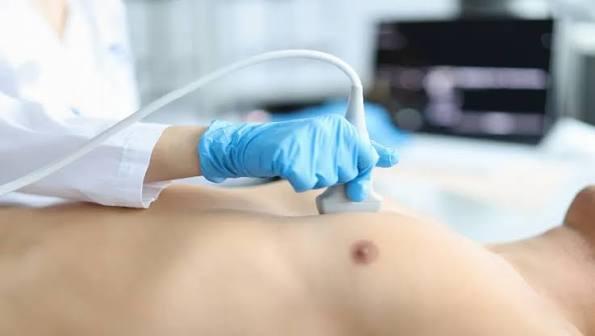
Echocardiography is the imaging of the heart using ultrasound. It provides detailed information about the heart's chambers, pumping function, valves, blood flow, and any potential structural problems.
- 20-40 Minutes
- Test Duration
- Non-invasive
- Method
- Same Visit
- Result Time
Who Needs Echocardiography, and When?
Because it directly answers questions about the heart's structure and function, echocardiography is one of the methods frequently used in cardiology assessment. The situations below are example scenarios in which this test may be considered; the final decision depends on the patient's individual examination.
Shortness of Breath or Reduced Exercise Capacity
It may be requested for complaints of increasing fatigue or shortness of breath during daily activities, to evaluate the heart's pumping function and chamber structure.
A Heart Murmur Is Heard
In patients found to have a heart murmur on examination, it may be used to investigate the source of the murmur and the structure of the valves.
Follow-up of Known Heart Failure or Valve Disease
In patients previously diagnosed with heart failure or valve disease, it may be repeated at regular intervals to monitor the course of the condition.
A Suspicious Finding Is Detected on the ECG
When a finding on the ECG suggests a possible structural problem, it may be requested to image the heart for a more detailed evaluation.
Difference Between Echocardiography (Transthoracic) and Transesophageal Echocardiography (TEE)
Transthoracic Echocardiography (Standard Echo)
Performed non-invasively through the chest wall; it provides a first-line assessment of the heart's structure and function.
Transesophageal Echocardiography (TEE)
Performed by advancing a probe into the esophagus; it provides much more detailed imaging from a position closer to the heart, and is preferred in specific situations where standard echo is insufficient.
What Does Echocardiography Cover?
Echocardiography provides a broad assessment of the heart's structure and function.
Included
- Measurement of the size and wall thickness of the heart chambers (atria and ventricles)
- Calculation of the heart's pumping function (ejection fraction)
- Evaluation of the structure and function of the heart valves (stenosis, regurgitation)
- Examination of the direction and speed of blood flow within the heart using Doppler
- Checking for fluid accumulation in the pericardium (the membrane surrounding the heart)
- Screening for findings associated with congenital heart anomalies
Not included
- Echocardiography does not directly image narrowing in the coronary arteries; coronary angiography or CT coronary angiography is used for this purpose.
- It does not continuously monitor the heart's electrical rhythm; an ECG or Holter ECG is required for rhythm assessment.
- In cases where standard (transthoracic) echo cannot provide sufficient image quality, transesophageal echocardiography (TEE) is considered as a separate procedure for more detailed examination.
Assessment and Approach
Correct interpretation of echocardiography results requires standard measurement protocols to be assessed together with the clinical context.
Standard Imaging Protocol
Care is taken to image the heart from different angles and standard windows, aiming for a complete assessment.
Comparison of Measurements with Reference Values
Measurements such as chamber size, wall thickness, and pumping function are compared with reference ranges appropriate for age and body size.
Holistic Assessment with Medical History
Imaging findings are interpreted together with the patient's complaints and prior cardiac history.
Determining the Need for Follow-up
Depending on the nature of the finding, it is evaluated whether repeat echo at regular intervals is needed.
How Is Echocardiography Performed?
Echocardiography follows a specific sequence and is a comfortable process for the patient.
- 01
Before the Test
Preparation
The patient exposes the upper body and lies on the examination table on the left side; no special preparation is required.
- 02
Start of the Test
Placement of the Probe
Ultrasound gel is applied to the chest, and the probe is moved over the chest from different angles.
- 03
During the Test
Imaging
The heart chambers, valves, and blood flow are imaged and recorded from different windows.
- 04
After the Test
Assessment
The images obtained are evaluated by the physician, and the measurements are interpreted and shared with the patient.
Factors That Affect the Assessment
The scope of the echocardiography assessment is shaped by individual clinical factors such as those below.
Presenting Complaint
The complaint the patient presents with — such as shortness of breath, a murmur, or palpitations — determines the areas the examination will focus on.
Known History of Heart Disease
A previously diagnosed valve disease, heart failure, or congenital heart anomaly affects the assessment.
Factors Affecting Image Quality
Factors such as body build and lung aeration can affect image quality; additional imaging methods are considered when needed.
Previous Echo Results
Any previous echocardiography recordings are included in the current assessment to track changes over time.
The information here is for general informational purposes only; the diagnosis and treatment plan are determined based on an individual assessment.
Frequently asked questions about Diagnostic Cardiology
Echocardiography is a painless, non-invasive diagnostic method in which the heart is imaged using ultrasound waves. It provides detailed information about the heart's chambers, pumping function, valves, and blood flow.
It is performed to evaluate the heart's structure and function when investigating complaints such as shortness of breath, a murmur, or palpitations, or when following up known heart failure or valve disease.
It may be recommended if you have complaints such as shortness of breath, fatigue, or palpitations, or if a murmur was detected on examination, in order to assess the heart's structure. This recommendation is based on the individual assessment your physician will make with you.
It generally does not require any special preparation. It is sufficient to wear clothing that can be easily removed from the upper body before the procedure.
The procedure generally takes between 20 and 40 minutes; the duration may vary depending on the scope of the findings to be evaluated.
No. Standard echocardiography is performed through the chest wall, while transesophageal echocardiography (TEE) is performed by advancing a probe into the esophagus, providing much more detailed imaging from a position closer to the heart.
Get in touch with us for an evaluation
Send your appointment request; we'll let you know about the evaluation process suited to your complaint and needs.