
Electrophysiology Study – What Is EPS?
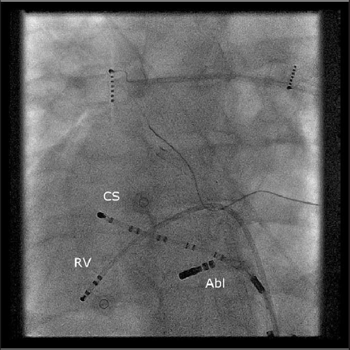
Electrophysiology study (EPS) is a specialized procedure performed to examine the heart's electrical system in detail. Thin catheters are typically advanced to the heart through vessels in the groin. Through these catheters, electrical signals from different regions of the heart are recorded directly.
- Local Anesthesia
- Anesthesia Type
- Catheter-Based
- Method
- Usually Same-Day
- Hospital Stay
Who Needs an Electrophysiology Study (EPS), and When?
EPS may be considered in various clinical situations where answers are sought about the heart's electrical system.
Those with unexplained palpitations
May be considered in patients with recurring palpitations whose surface ECG or Holter recordings show no clear findings, in order to clarify the source of the rhythm.
Those with a history of unexplained fainting (syncope)
Since fainting episodes may be related to an electrical rhythm disorder, evaluation may be undertaken in situations where the underlying mechanism needs to be investigated.
Those with suspected supraventricular tachycardia (such as AVNRT, WPW)
In cases with pre-excitation findings on ECG or a clinical picture suggestive of supraventricular tachycardia, it may be performed to reveal the mechanism of the rhythm.
Patients requiring mapping before catheter ablation
If a therapeutic ablation is being considered, electrical mapping with EPS may first be needed to identify the target area.
How does EPS compare with other methods?
Surface ECG and Holter
These recordings show the heart's rhythm externally, from the skin; EPS instead records signals directly from inside the heart via catheters, revealing the source of the rhythm disorder in more detail.
Catheter Ablation
EPS is primarily a diagnostic mapping procedure; if the findings are considered suitable, Catheter Ablation may be performed in the same session or in a separate session.
Echocardiography
Echocardiography provides structural and functional imaging of the heart; EPS examines the heart's electrical conduction system and the source of the rhythm.
What Does EPS Evaluate?
With EPS:
Included
- The source of the rhythm disorder causing palpitations
- Abnormal electrical pathways in the heart
- Electrical conduction problems
- The risk of certain rhythm disorders
Not included
- When necessary, catheter ablation can be performed during the same procedure as the electrophysiology study.
Evaluation and approach
A patient-specific evaluation process is followed before and during EPS.
Detailed history and record review beforehand
Before the procedure is planned, the patient's symptom history along with existing findings such as ECG and Holter recordings are reviewed together.
Continuous rhythm monitoring during the procedure
Once the catheters are positioned, the heart rhythm and electrical signals are monitored without interruption throughout the procedure.
Findings reviewed together with the patient
The electrophysiological findings obtained are interpreted together with the patient's clinical picture to clarify the next steps.
Close observation after the procedure
The catheter access site and the patient's general condition are closely monitored before discharge.
How Long Does the Procedure Take?
The procedure usually takes 30 minutes, and patients are mostly discharged the same day.
What clinical factors determine suitability?
Whether EPS is appropriate is assessed not by a single criterion but by looking at the patient as a whole. The evaluation generally takes the following factors into account.
Symptom history and current test results
The frequency and duration of complaints such as palpitations or syncope, along with findings from previous ECG/Holter recordings, form the basis of the evaluation.
Medications used
Whether blood thinners and rhythm-regulating medications in particular need to be adjusted before the procedure is also reviewed.
Accompanying structural heart disease
Echocardiographic findings such as heart valve structure and heart muscle function may affect how the procedure is planned.
General health status
Age, kidney function, and other accompanying health issues are evaluated together in terms of the timing and approach of the procedure.
The information here is for general informational purposes; the diagnosis and treatment plan are determined following an individualized evaluation.
Frequently asked questions about Interventional Electrophysiology
Electrophysiology study (EPS) is an interventional diagnostic method that directly examines the heart's electrical system using catheters. Thin catheters, introduced through a vessel in the groin and advanced to the heart, record the electrical signals within the heart and, when necessary, deliver controlled electrical stimuli to show how the rhythm disorder arises. This way, details that surface ECG cannot detect can be revealed.
EPS is performed to clarify the source of findings such as palpitations, unexplained fainting, or rhythm irregularities detected on ECG/Holter. The procedure reveals which region of the heart the rhythm disorder originates from, by what mechanism it is sustained, and how the conduction system is functioning. The findings obtained form the basis for determining whether follow-up or an additional procedure such as catheter ablation is needed.
EPS generally comes up in patients with unexplained palpitations, recurrent syncope, pre-excitation findings on ECG (such as WPW), or a suspected history of supraventricular/ventricular tachycardia. Whether the recommendation is made is determined by examining the person's symptoms, previous test results, and overall cardiology evaluation together; this decision is individualized and not based on a general rule.
The procedure is usually performed under local anesthesia and, when necessary, mild sedation; electrode catheters are introduced through a vessel in the groin and advanced to the heart under fluoroscopy guidance. The patient may be awake during catheter placement, and no significant pain is expected beyond a local anesthetic sensation; a feeling of palpitations may occur during the controlled electrical stimuli delivered through the catheters — this is a natural part of the procedure.
After the procedure is completed, brief pressure is applied over the catheter access site and the patient is kept under observation for a while. Most patients can be discharged the same day, although the observation period may vary depending on the findings or whether an additional procedure was performed. The results are reviewed together with the patient, and any next steps are clarified during this discussion.
No, the two serve different purposes. EPS is a diagnostic mapping procedure that records the heart's electrical activity and aims to identify the source of the rhythm disorder; catheter ablation is a separate intervention that, based on these findings, aims to treat the problematic tissue. When the findings are considered suitable, ablation can be performed in the same session or in a separate session.
Get in touch with us for an evaluation
Send your appointment request; we'll let you know about the evaluation process suited to your complaint and needs.