
TEE – What Is Transesophageal Echocardiography?
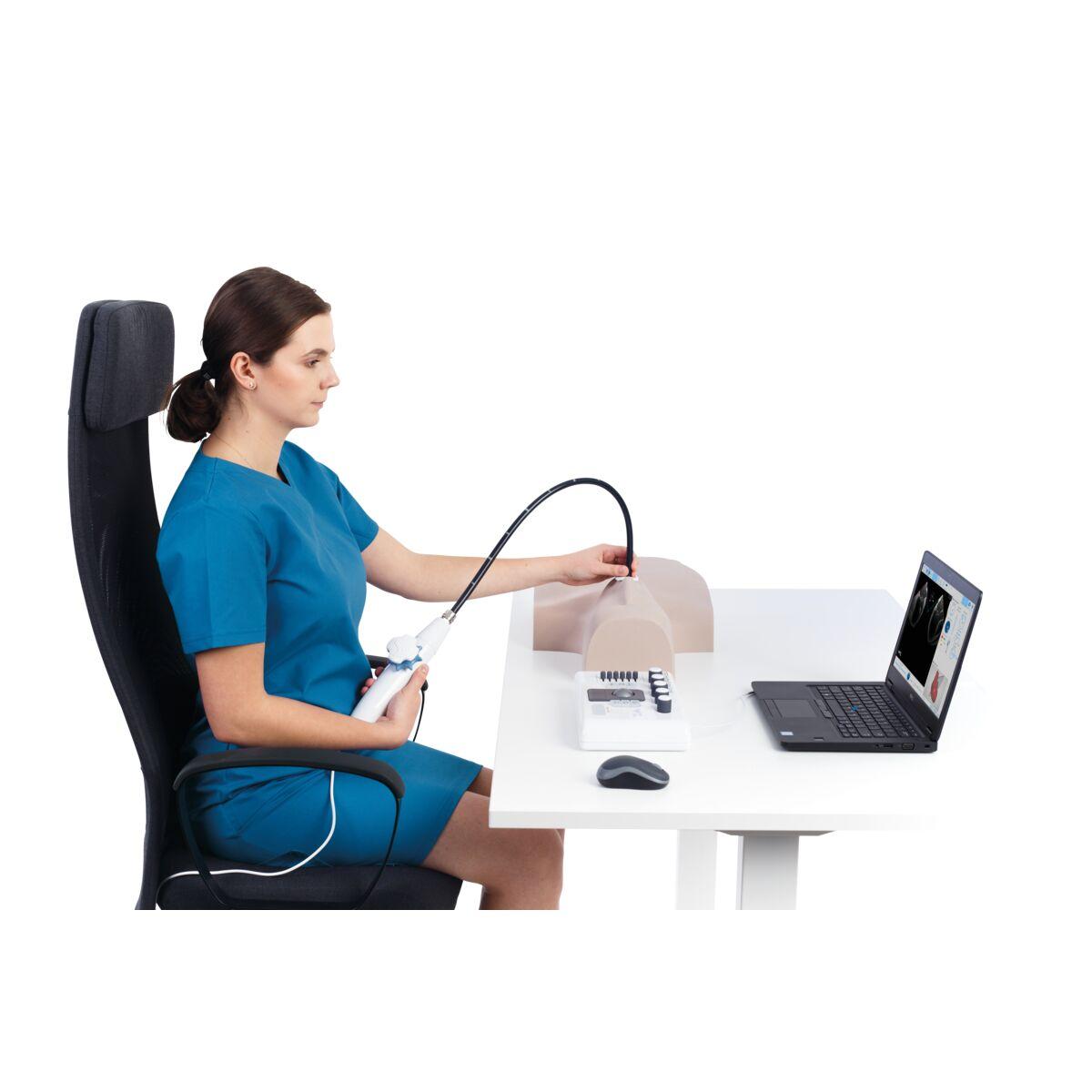
TEE is a specialized ultrasound method that provides more detailed imaging of the heart and heart valves. In this procedure, a thin probe with an ultrasound device at its tip is advanced into the esophagus. Because the heart lies directly in front of the esophagus, certain parts of the heart can be imaged in much greater detail than with standard echo.
- 20-40 Minutes
- Procedure Duration
- Light Sedation
- Method
- Short Observation Period
- After the Procedure
Who Needs TEE, and When?
TEE may be considered when standard echocardiography cannot provide sufficient detail, or when certain structures of the heart need to be visualized much more clearly. The situations below are example scenarios in which this procedure may be considered; the final decision depends on the patient's individual examination.
Situations Requiring Detailed Assessment of the Heart Valves
It may be requested when a suspicious finding involving the heart valves is detected on standard echo, in order to examine the valve structure in much greater detail.
Suspected Blood Clot in the Heart
It may be performed, for example, before certain procedures, particularly in patients with a rhythm disorder (such as atrial fibrillation), to investigate whether there is a blood clot in the heart.
Suspected Endocarditis (Heart Valve Infection)
It may be considered in patients suspected of having a heart valve infection, to visualize findings of the infection much more clearly than with standard echo.
Pre-Procedure Assessment Before TAVI or Catheter-Based Valve Repair
It may be performed in patients scheduled for interventional procedures such as TAVI or catheter-based heart valve repair, for detailed anatomical assessment before the procedure.
Difference Between TEE and Standard (Transthoracic) Echocardiography
Transesophageal Echocardiography (TEE)
Performed by advancing a probe into the esophagus; it provides much more detailed imaging from a position much closer to the heart, particularly for the valves and structures behind the heart.
Standard (Transthoracic) Echocardiography
Performed non-invasively through the chest wall; it provides a first-line assessment of the heart's general structure and function and is sufficient in most cases.
Which Conditions Is TEE Used to Assess?
TEE is used in particular to assess the following:
Included
- Heart valves
- Blood clots within the heart
- Openings in the heart, such as ASD and PFO
- Heart valve infection (endocarditis)
- Structural heart disease
- Pre-procedure assessment before interventions such as TAVI and catheter-based heart valve repair
Not included
Assessment and Approach
Because TEE is a more specialized procedure than standard echo, specific assessment steps are followed to ensure safety and an accurate result.
Pre-Procedure Suitability Assessment
Whether there are any issues related to the esophagus or stomach is assessed before the procedure.
Performed on an Empty Stomach
To reduce the risk of aspiration, the procedure is performed after the patient has fasted for a specific period.
Monitoring During Sedation
In patients given light sedation, oxygen level and general condition are closely monitored throughout the procedure.
Post-Procedure Observation
After the procedure, the patient is kept under short-term observation, particularly until the effects of sedation wear off.
How Is TEE Performed?
TEE is a planned procedure that includes specific preparation, imaging, and observation phases.
- 01
Before the Procedure
Pre-Assessment and Preparation
The patient is required to fast, and any issues related to the esophagus are reviewed.
- 02
Start of the Procedure
Numbing the Throat
The throat area is numbed with local anesthesia, and light sedation is given if needed.
- 03
During the Procedure
Imaging
The ultrasound probe is gently advanced into the esophagus, and the heart is imaged in detail from different angles.
- 04
After the Procedure
Observation and Assessment
The probe is removed and the patient is kept under observation for a short period; the images obtained are evaluated by the physician and shared with the patient.
Factors That Affect the Assessment
The suitability and timing of TEE are determined by taking into account individual clinical factors such as those below.
Reason for Referral
The purpose for which it is requested — such as valve assessment, investigation for a clot, or pre-procedure planning — determines the scope of the assessment.
History Related to the Esophagus and Stomach
Whether there is a known issue related to the esophagus is taken into account when planning the procedure.
Standard Echo Results
The results of a previously performed transthoracic echo and any findings requiring clarification are included in the assessment.
General Health Status
In patients who will receive sedation, general health status and any accompanying conditions are taken into account during the assessment process.
The information here is for general informational purposes only; the diagnosis and treatment plan are determined based on an individual assessment.
Frequently asked questions about Diagnostic Cardiology
TEE is a specialized ultrasound method in which a thin device with an ultrasound probe at its tip is advanced into the esophagus, allowing the heart to be imaged in much greater detail.
It is performed for detailed assessment of the heart valves, investigation of a blood clot in the heart, examination of a structural opening such as ASD/PFO or suspected endocarditis, or pre-procedure assessment before interventions such as TAVI.
It may be recommended if there is a finding on standard echocardiography that could not be clarified, if a heart valve infection is suspected, or if detailed assessment is needed before an interventional procedure. This recommendation is based on the individual assessment your physician will make with you.
During the procedure, the throat is numbed with local anesthesia, and light sedation is given when needed; as a result, the procedure is well tolerated by most patients.
Yes. To reduce the risk of aspiration, fasting for a specific period is required before the procedure; this period is specified by the physician.
No. Standard echo is performed through the chest wall, while TEE is performed by advancing a probe into the esophagus, providing much more detailed imaging from a position much closer to the heart, particularly for the valves.
Get in touch with us for an evaluation
Send your appointment request; we'll let you know about the evaluation process suited to your complaint and needs.